


Home›The Treatise›Chapters›Chapter 22
Tratado de Cirurgia da Coluna Vertebral
SECTION 3 • Traumatic Spine Injuries
Chapter22

Traumatic Injuries of the Thoracic and Lumbar Spine

Vancouver: Jaccard APB, Rodriguez CAA, Vieira MB📖 Pages: 319-330
Full reading of this chapter is available exclusively in the printed edition of the Treatise.
Sec. 3Traumatic Spine Injuries
Cap. 22Clinical Chapter
3authors
Português
Español
English
Referênciasscientific citations
📑

Chapter Summary

• Context: Traumatic injuries of the thoracic and lumbar spine represent the most frequent fractures of the entire vertebral axis, predominantly concentrating across the biomechanically vulnerable thoracolumbar junction (T10–L2). High-energy trauma (motor vehicle collisions, falls from height) affects younger populations with substantial risk of conus medullaris and cauda equina injury, whereas low-energy fragility fractures affect osteoporotic elderly individuals. The functional spinal unit comprises the anterior column (vertebral bodies, intervertebral discs, anterior and posterior longitudinal ligaments) and the posterior tension band (pedicles, lamina, facet joints, and the Posterior Ligamentous Complex [PLC: supraspinous, interspinous ligaments, ligamentum flavum, and facet capsules]). Classifications have evolved from mechanistic models (Denis three-column model, McCormack Load Sharing Classification) to the globally standardized AO Spine Thoracolumbar Classification and Thoracolumbar Injury Classification and Severity Score (TLICS). Multi-detector CT defines fracture morphology, comminution, and canal compromise, while MRI evaluates PLC disruption, cord contusion, and epidural hematoma. The central therapeutic challenge is distinguishing mechanically stable compression injuries amenable to functional bracing from unstable burst, distraction (Chance), or rotational translation fractures requiring open or percutaneous posterior pedicle screw instrumentation, anterior column cage reconstruction, or spinal decompression.
• Chapter Objective: To present the clinical evaluation, classification, biomechanical stability assessment, and non-operative/surgical management of thoracic and lumbar spine trauma. The reader should be able to apply the AO Spine Thoracolumbar Classification (Types A, B, C) and the TLICS scoring system; recognize the clinical and radiological hallmarks of posterior ligamentous complex (PLC) disruption; understand the indications for percutaneous versus open pedicle screw fixation, direct vs. indirect decompression, and anterior column reconstruction; and prevent post-traumatic kyphosis and neurological deterioration.
• Biomechanics and the Thoracolumbar Transition ZoneThe thoracolumbar junction (T10–L2) transitions from the rigid thoracic cage (coronal facets, kyphosis) to the mobile lumbar spine (sagittal facets, lordosis). Denis established the 3-column model (anterior, middle, posterior). Mechanical stability relies fundamentally on the integrity of the middle column (posterior vertebral body cortex and PLL) and the posterior tension band (PLC). Loss of middle column integrity (burst fracture) combined with PLC disruption results in progressive mechanical collapse and post-traumatic kyphotic deformity.
• AO Spine Thoracolumbar Classification SystemThe AO Spine system organizes injuries hierarchically into three primary morphological types: 1) Type A: Compression fractures (A0 minor non-structural, A1 wedge compression, A2 coronal split/pincer, A3 incomplete burst involving one endplate, A4 complete burst involving both endplates and posterior wall retropulsion); 2) Type B: Distraction injuries / Tension band failure (B1 transosseous Chance fracture through posterior elements and vertebral body, B2 osseoligamentous posterior tension band disruption, B3 hyperextension injury through anterior tension band/ALL); 3) Type C: Translation/Displacement in any plane (subluxation, dislocation, fracture-dislocation). Neurological status is classified from N0 (intact) to N4 (complete cord/conus deficit), and clinical modifiers include M1 (indeterminate PLC) and M2 (comorbidities/ankylosing spine).
• TLICS Score and Decision-Making AlgorithmsThe Thoracolumbar Injury Classification and Severity Score (TLICS) assigns points across three core categories: 1) Morphology (compression = 1, burst = 2, distraction = 3, translation/rotation = 4); 2) Neurological status (intact = 0, nerve root injury = 2, complete cord = 2, incomplete cord/conus = 3, cauda equina syndrome = 3); 3) PLC integrity (intact = 0, suspected/indeterminate = 2, disrupted = 3). A total TLICS score <=3 indicates non-operative management (early mobilization with or without a rigid TLSO brace); TLICS = 4 is indeterminate (surgeon discretion); and TLICS >=5 mandates surgical reduction and stabilization.
• Diagnostic Imaging and Clinical AssessmentMulti-detector CT with multiplanar reconstructions is the primary investigation to assess vertebral body height loss, sagittal kyphosis (Cobb angle), pedicle splay, and canal stenosis. MRI (STIR and T2-weighted sequences) is mandatory when physical examination reveals focal interspinous widening or palpation tenderness, demonstrating PLC hyperintensity (black line disruption of ligamentum flavum/supraspinous ligament). MRI also evaluates conus medullaris contusion and traumatic disc disruption.
• Management Principles: Conservative, Percutaneous, and Open ReconstructionNon-operative treatment (early ambulation, Jewett or molded TLSO brace, serial standing radiographs at 1, 2, and 6 weeks) is indicated for stable A1, A2, and neurologically intact A3 fractures with intact PLC and local kyphosis <15°–20°. Surgical treatment is indicated for unstable burst fractures (A4 with significant comminution), distraction injuries (B1, B2, B3), fracture-dislocations (Type C), or any fracture with progressive neurological deficit. Surgical options include minimally invasive percutaneous pedicle screw fixation (sparing paraspinal muscles in intact neurology), open posterior decompression and instrumentation with intermediate screws, and anterior or posterolateral corpectomy with expandable cage reconstruction for severe anterior load-bearing deficiency (McCormack Load Sharing score >=7).
• Clinical Application: In clinical practice, evaluate three interconnected factors: neurological deficit, PLC integrity, and anterior column comminution. In a patient with an L1 burst fracture, intact neurology, and intact PLC on MRI (TLICS 2), non-operative treatment with a molded TLSO brace and immediate protected ambulation yields functional outcomes equivalent to open surgery without operative morbidity. In contrast, if MRI reveals hyperintensity of the supraspinous ligament and ligamentum flavum (PLC disruption, AO Type B2 / TLICS 5), surgery is mandatory even in a neurologically intact patient, as non-operative bracing fails, leading to progressive kyphosis and chronic back pain. In patients with an A4 burst fracture and progressive neurological deficit from retropulsed bone fragments, urgent posterior decompression, postural ligamentotaxis reduction, and pedicle screw fixation within 24 hours provide excellent canal clearance and neurological recovery. In patients with high McCormack Load Sharing scores (>=7) undergoing short-segment posterior instrumentation, anterior column support (interbody cage) is necessary to prevent posterior hardware fatigue failure.
🏷️

Keywords

Preferred DeCS/MeSH Descriptors:
Thoracic VertebraeLumbar VertebraeSpinal FracturesSpinal Cord InjuriesKyphosisTomography, X-Ray ComputedMagnetic Resonance ImagingSpinal Fusion
⭐

Why this chapter matters

A thoracolumbar fracture can appear to be a benign compression fracture on standard X-rays while harboring an unrecognized complete posterior ligamentous complex (PLC) rupture that will progress to severe post-traumatic kyphosis and chronic disability if treated in a simple brace. This chapter provides a rigorous diagnostic framework combining clinical examination, multi-detector CT, and MRI to identify occult tension band failures, calculate TLICS and AO scores, and select the least invasive, most durable stabilization strategy.

“Accurate diagnosis and management of thoracic and lumbar spine trauma depend on identifying the primary injury mechanism and evaluating true structural stability rather than relying solely on bony fracture appearance. The AO Spine classification and TLICS score integrate morphology, posterior ligamentous complex (PLC) competence, and neurological status. Selecting between conservative functional bracing, percutaneous instrumentation, and open anterior/posterior reconstruction must be tailored to patient stability, neurology, and anterior column load sharing.”
✨

Chapter Highlights

🌐
Card 1 — Core Concept
PLC Integrity Governs Stability

The Posterior Ligamentous Complex (PLC: supraspinous, interspinous, ligamentum flavum, facet capsules) prevents progressive kyphotic collapse. Any fracture with PLC disruption (AO Type B / TLICS >=5) is mechanically unstable and mandates surgical stabilization.

🩺
Card 2 — Clinical Decision
TLICS Score Determines Treatment Strategy

Apply the TLICS algorithm: score <=3 indicates non-operative bracing with early mobilization; score = 4 is indeterminate; score >=5 requires surgical stabilization (percutaneous or open instrumentation).

📐
Card 3 — Key Pearl / Warning
Recognize Occult Distraction Injuries

Always evaluate spinous process widening and MRI STIR hyperintensity. What appears as a simple anterior wedge fracture on CT can represent a severe flexion-distraction injury (AO B2) if the posterior tension band is torn.

📑

How to Cite this Chapter (Vancouver Format)

Official bibliographic indexing and citation guidelines
📖 Pages: 319-330Vancouver Style
Authors (Vancouver):Jaccard APB, Rodriguez CAA, Vieira MB

Jaccard APB, Rodriguez CAA, Vieira MB. Lesões traumáticas da coluna torácica e lombar. In: Pudles E, Defino H, Risso M, editors. Tratado de Cirurgia da Coluna Vertebral (Treatise of Spine Surgery). 1st ed. Rio de Janeiro: Dilivros Editora; 2026. p. 319-330.

ISBN: 978-85-8053-292-0 • 1.ª Edição • Dilivros Editora
📚

Bibliographic References

1. Bezerra Junior DL, Sá BFA, Pokorny GHO, Almeida NS, Silva DRC, Silva JPG, et al. Epidemiological analysis of patients victims of surgical thoracic/lumbar fractures treated at a tertiary hospital in Brazil. J Bras Neurocirur. 2023;34(4):422-7. doi:10.22290/jbnc.2023.340402.
2. National Spinal Cord Injury Statistical Center. Spinal cord injury facts and figures at a glance. Birmingham, AL: University of Alabama at Birmingham; 2021.
3. Cunha FM, Menezes CM, Guimarães EP. Lesões traumáticas da coluna torácica e lombar. Rev Bras Ortop. 2000;35(1/2):17-22.
4. Nordin M, Frankel VH. Biomechanics of the lumbar spine. In: Basic biomechanics of the musculoskeletal system. 4th ed. Philadelphia: Lippincott Williams & Wilkins; 2012. p. 255-85.
5. Gamanagatti S, Rathinam D, Rangarajan K, Kumar A, Farooque K, Sharma V. Imaging evaluation of traumatic thoracolumbar spine injuries: radiological review. World J Radiol. 2015;7(9):253-65. doi:10.4329/wjr.v7.i9.253.
6. Leone A, Guglielmi G, Cassar-Pullicino VN, Bonomo L. Lumbar intervertebral instability: a review. Radiology. 2007;245(1):62-77. doi:10.1148/radiol.2451051359.
7. Reinhold M, Knop C, Beisse R, Audigé L, Kandziora F, Pizanis A, et al. Operative treatment of traumatic fractures of the thoracic and lumbar spine: Part I — Epidemiology. Eur Spine J. 2010;19(7):1257-69. doi:10.1007/s00586-010-1580-2.
8. Ghezelbash F, Alizadeh M, Ahmadian A, Gholami M, Rahimi M, Karami M, et al. The role of the thoracolumbar junction in spinal biomechanics: a finite element study. J Orthop Surg Res. 2022;17(1):335. doi:10.1186/s13018-022-02954-6.
9. Wood KB, Li W, Lebl DS, Ploumis A. Management of thoracolumbar spine fractures. Spine J. 2014;14(1):145-64. doi:10.1016/j.spinee.2012.10.041.
10. VandenBerg J, Cullison K, Fowler SA, Parsons MS, McAndrew CM, Carpenter CR. Blunt thoracolumbar-spine trauma evaluation in the emergency department: a meta-analysis of diagnostic accuracy for history, physical examination, and imaging. J Emerg Med. 2019;56(2):153-65. doi:10.1016/j.jemermed.2018.10.032.
11. Maynard FM Jr, Bracken MB, Creasey G, Ditunno JF Jr, Donovan WH, Ducker TB, et al. International standards for neurological and functional classification of spinal cord injury. Spinal Cord. 1997;35(5):266-74.
12. Gerges C, Raghavan A, Wright J, Shammassian B, Wright CH, Moore T. Cervical, thoracolumbar, and sacral spine trauma classifications: past, present, and future. Neurol Res. 2020;42(10):857-66. doi:10.1080/01616412.2020.1797373.
13. Schnake KJ, Schroeder GD, Vaccaro AR, Oner C. AOSpine Classification Systems (Subaxial, Thoracolumbar). J Orthop Trauma. 2017;31 Suppl 4:S14-S23. doi:10.1097/BOT.0000000000000947.
14. Yelamarthy PKK, Chhabra HS, Vaksha V, Agarwal Y, Agarwal A, Das K, et al. Radiological protocol in spinal trauma: literature review and Spinal Cord Society position statement. Eur Spine J. 2020;29(6):1197-1211. doi:10.1007/s00586-019-06112-z.
15. Izzo R, Al Qassab S, Popolizio T, Balzano RF, Perri M, Cassar-Pullicino V, et al. Imaging of thoracolumbar spine traumas. Eur J Radiol. 2022;154:110343. doi:10.1016/j.ejrad.2022.110343.
16. Verheyden AP, Spiegl UJ, Ekkerlein H, Gercek E, Hauck S, Josten C, et al. Treatment of fractures of the thoracolumbar spine: recommendations of the Spine Section of the German Society for Orthopaedics and Trauma (DGOU). Global Spine J. 2018;8(2 Suppl):34S-45S. doi:10.1177/2192568218771668.
17. Spiegl UJ, Fischer K, Schmidt J, Schnoor J, Delank S, Josten C, et al. The conservative treatment of traumatic thoracolumbar vertebral fractures. Dtsch Arztebl Int. 2018;115(42):697-704. doi:10.3238/arztebl.2018.0697.
18. Kweh BTS, Tee JW, Dandurand C, Vaccaro AR, Lorin BM, Schnake K, et al. The AO Spine Thoracolumbar Injury Classification System and treatment algorithm in decision making for thoracolumbar burst fractures without neurologic deficit. Global Spine J. 2024;14(1 Suppl):32S-40S. doi:10.1177/21925682231195764.
19. Nau C, Pape HC, Jug M, Wendt K, Komadina R, Bloemers F. Thoracolumbar injuries: non operative treatment: indications, management. Eur J Trauma Emerg Surg. 2024;50(5):1951-1957. doi:10.1007/s00068-024-02619-3.
20. Yuan H, Chang QY, Chen J, Wang YT, Gan ZJ, Wen S, et al. A retrospective analysis of the effects of different analgesics on the pain of patients with traumatic thoracolumbar fractures in the peri-treatment period. J Orthop Surg Res. 2021;16(1):268. doi:10.1186/s13018-021-02401-w.
21. Rosenthal BD, Boody BS, Jenkins TJ, Hsu WK, Patel AA, Savage JW. Thoracolumbar burst fractures. Clin Spine Surg. 2018;31(4):143-151. doi:10.1097/BSD.0000000000000634.
22. Urquhart JC, Alrehaili OA, Fisher CG, Fleming A, Rasoulinejad P, Gurr K, et al. Treatment of thoracolumbar burst fractures: extended follow-up of a randomized clinical trial comparing orthosis versus no orthosis. J Neurosurg Spine. 2017;27(1):42-47. doi:10.3171/2016.11.SPINE161031.
23. Linhares D, Pinto BS, Ribeiro da Silva M, Neves N, Fonseca JA. Orthosis in thoracolumbar fractures: a systematic review and meta-analysis of randomized controlled trials. Spine (Phila Pa 1976). 2020;45(22):E1523-E1531. doi:10.1097/BRS.0000000000003655.
24. Bloemers F, Jug M, Nau C, Komadina R, Pape HC, Wendt K. Thoracolumbar injuries: operative treatment: indications, techniques, timing and implant removal. Current practice. Eur J Trauma Emerg Surg. 2024;50(5):1959-1968. doi:10.1007/s00068-024-02602-y.
25. Sharif S, Shaikh Y, Yaman O, Zileli M. Surgical techniques for thoracolumbar spine fractures: WFNS Spine Committee recommendations. Neurospine. 2021;18(4):667-680. doi:10.14245/ns.2142206.253.
26. Giorgi PD, Legrenzi S, Youchen Y, Federico B, Schirò GR. The effectiveness of posterior Schanz screw system in the anatomical restoration of thoracolumbar vertebral burst fractures: a 10-year single-institution experience. J Craniovertebr Junction Spine. 2024;15(4):411-418. doi:10.4103/jcvjs.jcvjs_118_24.
27. AO Foundation. Surgery Reference: posterior short segment fixation with Schanz pins. https://surgeryreference.aofoundation.org/spine/trauma/thoracolumbar/b2/posterior-short-segment-fixation-with-schanz-pins
28. Esposito F, Bove I, Vitulli F, Bocchino A, Barbanera A, Nape S, et al. Outcome measures of open versus minimally invasive surgery for thoracolumbar spinal traumatic fractures: a systematic review and meta-analysis. J Clin Med. 2024;13(18):5558. doi:10.3390/jcm13185558.
29. Soultanis K, Thano A, Soucacos PN. Outcome of thoracolumbar compression fractures following non-operative treatment. Injury. 2021;52(12):3685-3690. doi:10.1016/j.injury.2021.05.019.
30. Cabrera JP, Carazzo CA, Guiroy A, White KP, Guasque J, Sfreddo E, et al. Risk factors for postoperative complications after surgical treatment of type B and C injuries of the thoracolumbar spine. World Neurosurg. 2023;170:e520-e528. doi:10.1016/j.wneu.2022.11.059.
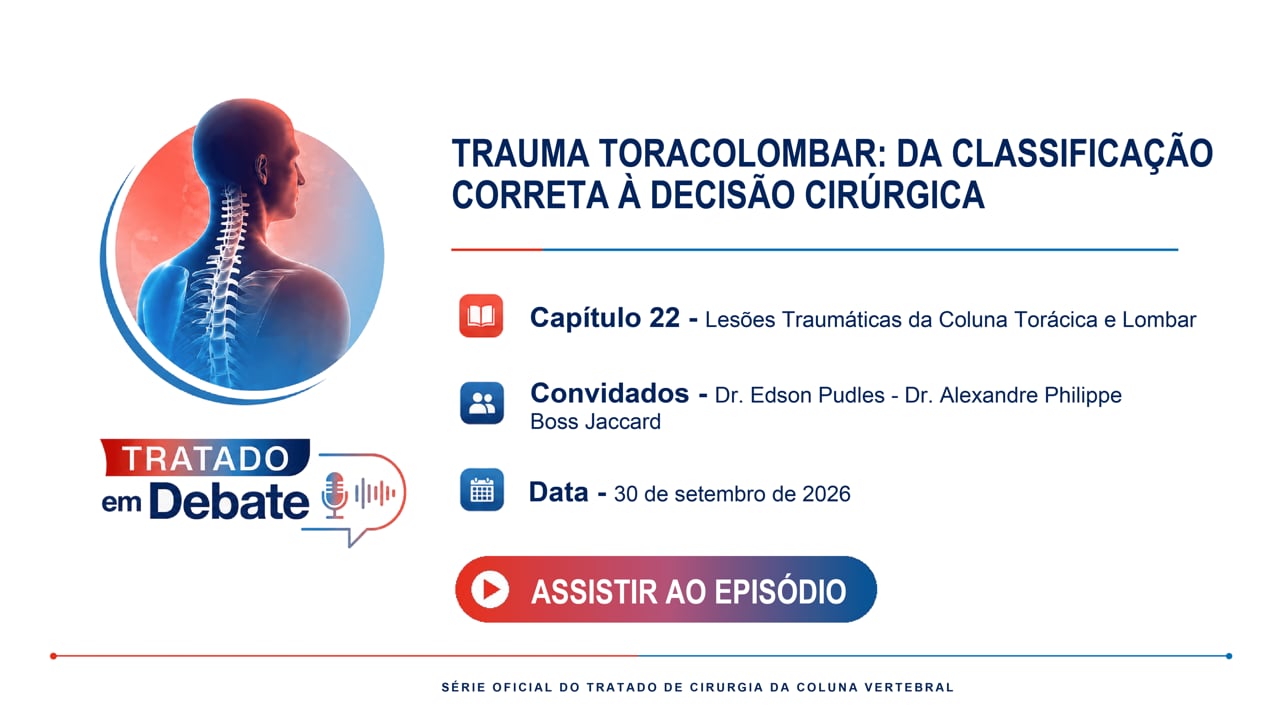
Episode 05 – Traumatic Injuries of the Thoracic and Lumbar Spine
Premiere Countdown
Exclusive Premiere • Wednesday, September 30 at 9:00 PM (BRT)
00Days
:
04Hours
:
46Mins
:
15Secs

The full videocast will premiere automatically in this player on Wednesday, 09/30 at 6:00 PM (BRT).

Also premiering on Spotify
Treatise in Debate

Official videocast derived from the treatise chapters.

Episode 05 – Traumatic Injuries of the Thoracic and Lumbar Spine

Thoracolumbar Trauma: from Proper Classification to Surgical Decision