


Home›The Treatise›Chapters›Chapter 14
Tratado de Cirurgia da Coluna Vertebral
SECTION 2 • Diagnosis
Chapter14

Intraoperative Neurophysiological Monitoring in Spine Surgery

Vancouver: Ferreira RJR, Nogueira PTB, Pontes RWF📖 Pages: 185-200
Full reading of this chapter is available exclusively in the printed edition of the Treatise.
Sec. 2Diagnosis
Cap. 14Clinical Chapter
3authors
Português
Español
English
Referênciasscientific citations
📑

Chapter Summary

• Context: Intraoperative neurophysiological monitoring (IONM) provides real-time functional assessment of the central and peripheral nervous system during spine surgery, serving as an early-warning diagnostic tool to detect impending iatrogenic neural injury, guide safe corrective maneuvers, and prevent postoperative neurological deficits. Multimodal IONM integrates transcranial motor evoked potentials (TcMEP), somatosensory evoked potentials (SSEP), spontaneous and triggered electromyography (sEMG and tEMG), and dermatomal/reflex responses (D-wave, bulbocavernosus reflex). Each modality monitors distinct neural pathways with specific temporal sensitivities and alarm criteria: SSEPs assess the dorsal column-medial lemniscal sensory pathway (supplied by the posterior spinal arteries), while TcMEPs evaluate the corticospinal motor pathway and anterior horn cells (supplied by the anterior spinal artery). Consequently, selective anterior spinal artery ischemia or direct anterior motor tract contusion can abolish TcMEPs without altering SSEPs. Free-run sEMG detects mechanical nerve root traction, irritation, or compression during instrumentation and decompression, while pedicle screw triggered EMG (tEMG) detects pedicle cortical breaches. Successful IONM requires total intravenous anesthesia (TIVA) without halogenated inhalational agents or neuromuscular blockade (which abolish MEPs and EMG), temperature and blood pressure optimization, and adherence to structured institutional checklists to identify and rapidly reverse intraoperative neurological insults.
• Chapter Objective: To present the neurophysiological principles, modalities, alarm criteria, and anesthetic protocols of multimodal intraoperative neuromonitoring (IONM) in spine surgery. The reader should be able to understand the physiological pathways evaluated by SSEP, TcMEP, EMG, D-wave, and reflex testing; apply multimodal monitoring in deformity, cervical myelopathy, intradural tumor, and minimally invasive lateral surgery; recognize significant neurophysiological alarm threshold criteria; and execute structured intraoperative rescue protocols following acute signal degradation.
• Multimodal IONM Modalities and Neural PathwaysMultimodal IONM combines complementary neurophysiological modalities: 1) Somatosensory Evoked Potentials (SSEP): elicited by peripheral nerve stimulation (median, ulnar, posterior tibial nerves) and recorded over the primary somatosensory cortex and cervical spine, evaluating the ascending dorsal column-medial lemniscal sensory pathways; 2) Transcranial Motor Evoked Potentials (TcMEP): generated by transcranial electrical stimulation over the motor cortex and recorded as compound muscle action potentials (CMAPs) from target limb muscles, evaluating the descending lateral corticospinal tracts and alpha motor neurons; 3) D-Wave: recorded directly from the spinal epidural space via a catheter electrode, evaluating direct pyramidal tract axonal conduction independent of neuromuscular junction transmission; 4) Spontaneous EMG (sEMG): continuous monitoring of root-specific muscles for neurotonic burst and train discharges indicating mechanical root irritation; 5) Triggered EMG (tEMG): delivering electrical current to pedicle screw probes to detect bone breaches; and 6) Bulbocavernosus Reflex (BCR): monitoring the S2–S4 conus medullaris and pudendal reflex arc.
• Validated Alarm Threshold CriteriaNeurophysiological alarm criteria define significant signal loss requiring immediate surgical and systemic action: 1) SSEP alarm: a >=50% decrease in amplitude and/or a >=10% increase in latency compared to stable baseline; 2) TcMEP alarm: a >=50–80% amplitude reduction, significant threshold voltage increase (>100 V), or complete loss of CMAP response in one or more limb muscles; 3) D-Wave alarm: a >=50% reduction in D-wave amplitude during intramedullary tumor resection indicates permanent motor tract injury; 4) sEMG alarm: neurotonic train activity (sustained high-frequency discharge) indicates active mechanical root stretch or compression; 5) Triggered EMG: pedicle screw threshold <6–8 mA in the lumbar spine or <5–6 mA in the thoracic spine indicates medial/inferior pedicle wall breach with direct nerve root contact.
• Anesthetic Requirements: TIVA ProtocolAnesthesia profoundly influences IONM signal acquisition. Halogenated volatile inhalational anesthetics (sevoflurane, desflurane, isoflurane) and nitrous oxide produce dose-dependent suppression of anterior horn alpha motor neurons, markedly blunting or abolishing TcMEP and H-reflex responses. Therefore, Total Intravenous Anesthesia (TIVA)—utilizing continuous infusions of propofol and short-acting opioids (remifentanil, sufentanil, fentanyl), with or without low-dose ketamine/dexmedetomidine—is mandatory. Neuromuscular blocking agents (paralytics) can be used for initial endotracheal intubation but must be completely avoided or reversed during monitoring (train-of-four [TOF] maintained at 4/4 twitches) to permit muscle MEP and EMG recordings.
• Clinical Indications Across Spine SubspecialtiesIONM indications are tailored to surgical risk: 1) Adult and adolescent deformity (scoliosis/kyphosis): continuous TcMEP and SSEP monitoring during correction maneuvers (derotation, translation, distraction) and osteotomies (PCO, PSO, VCR); 2) Cervical and thoracic myelopathy: TcMEP and SSEP monitoring from patient positioning (head positioning and traction) through decompression; 3) Intramedullary spinal cord tumors (ependymomas, astrocytomas): combined TcMEP, SSEP, and D-wave monitoring to establish safe resection margins (stopping resection if D-wave drops >50%); 4) Lateral lumbar interbody fusion (LLIF transpsoas): real-time triggered EMG directional mapping to detect and avoid lumbar plexus branches within the psoas; 5) Lumbosacral instrumented fusion: tEMG screw testing and BCR monitoring to protect roots and bowel/bladder function.
• Structured Intraoperative Rescue ProtocolUpon an IONM alert, a rapid, standardized multidisciplinary checklist must be executed simultaneously: 1) Systemic optimization: verify mean arterial pressure (MAP target >80–85 mmHg to restore cord perfusion), check core temperature (>35.5°C), hematocrit (>28–30%), arterial blood gases, and rule out anesthetic bolus or volatile gas accidental administration; 2) Technical verification: check recording/stimulating electrodes and machine grounding; 3) Surgical intervention: immediately halt active surgical maneuvers, remove compressive retractors, irrigate wound with warm saline, release temporary correction rods or distraction forces, reverse deformity correction, and remove recently placed screws or interbody cages. If signals fail to recover despite optimization, consider an intraoperative wake-up test (Stagnara wake-up test).
• Clinical Application: In spine deformity surgery, TcMEPs detect impending spinal cord ischemia or stretch injury minutes before SSEP changes appear. When a sudden loss of lower extremity MEPs occurs during scoliosis rod derotation or vertebral column resection (VCR), the surgical team immediately raises MAP to >85–90 mmHg, pauses the procedure, and releases rod tension. In over 85–90% of cases, timely release of correction and perfusion augmentation restore MEP signals and prevent permanent paraplegia. In thoracic and lumbar pedicle screw placement, triggered EMG with thresholds <6 mA accurately flags misplaced screws encroaching on the spinal canal or exiting nerve root, enabling immediate repositioning before wound closure. In intramedullary tumor resections, preserving a D-wave amplitude >50% ensures that the patient will maintain long-term ambulatory motor function, even if transient postoperative paresis occurs due to spinal cord stunning.
🏷️

Keywords

Preferred DeCS/MeSH Descriptors:
Intraoperative Neurophysiological MonitoringEvoked Potentials, MotorEvoked Potentials, SomatosensoryElectromyographySpinal Cord InjuriesSpineAnesthesia, IntravenousSurgical Procedures, Operative
⭐

Why this chapter matters

A postoperative neurological deficit is among the most devastating complications in spine surgery. IONM provides real-time feedback during the reversible window of neural compromise. Relying on SSEPs alone risks missing pure motor tract ischemia, while monitoring without TIVA results in uninterpretable signals. This chapter provides the evidence-based neurophysiological parameters, anesthetic guidelines, and immediate intraoperative response algorithms necessary to safeguard neural function across complex deformity, degenerative, tumor, and minimally invasive spine procedures.

“Multimodal intraoperative neuromonitoring (TcMEP, SSEP, EMG, D-wave) acts as a dynamic neurological examination in the anesthetized patient. Integrating motor and sensory modalities under total intravenous anesthesia (TIVA), adhering to standardized alarm criteria (>=50% SSEP drop, >=50–80% TcMEP loss, tEMG <6 mA), and executing structured multidisciplinary rescue protocols (MAP elevation >85 mmHg, release of surgical distraction) significantly minimizes catastrophic neurological complications in spine surgery.”
✨

Chapter Highlights

🌐
Card 1 — Core Concept
Multimodal Monitoring is Mandatory

SSEPs monitor dorsal columns (posterior spinal artery), while TcMEPs monitor corticospinal motor tracts (anterior spinal artery). Anterior cord ischemia or motor tract compression can cause complete paraplegia while SSEPs remain entirely normal. Always utilize combined TcMEP and SSEP monitoring.

🩺
Card 2 — Clinical Decision
Standardized IONM Alarm Thresholds

Recognize significant alarm criteria: >=50% drop in SSEP amplitude, >=50–80% loss of TcMEP amplitude, >=50% decrease in D-wave, sustained neurotonic sEMG trains, or pedicle screw triggered EMG threshold <6 mA in the lumbar spine (indicating medial cortical breach).

📐
Card 3 — Key Pearl / Warning
Immediate Rescue Algorithm

Upon acute IONM signal loss: immediately elevate Mean Arterial Pressure (MAP > 80–85 mmHg), rule out anesthetic boluses/paralytics, halt surgical manipulation, warm the surgical field, and release correction rods or distraction. Timely reversal restores signals and prevents permanent deficits.

📑

How to Cite this Chapter (Vancouver Format)

Official bibliographic indexing and citation guidelines
📖 Pages: 185-200Vancouver Style
Authors (Vancouver):Ferreira RJR, Nogueira PTB, Pontes RWF

Ferreira RJR, Nogueira PTB, Pontes RWF. Monitorização neurofisiológica intraoperatória em cirurgia de coluna. In: Pudles E, Defino H, Risso M, editors. Tratado de Cirurgia da Coluna Vertebral (Treatise of Spine Surgery). 1st ed. Rio de Janeiro: Dilivros Editora; 2026. p. 185-200.

ISBN: 978-85-8053-292-0 • 1.ª Edição • Dilivros Editora
📚

Bibliographic References

1. As referências foram mantidas na ordem e com a numeração apresentadas no capítulo. A pontuação e a disposição foram uniformizadas segundo o estilo Vancouver, sem completar por suposição informações ausentes.
2. Vauzelle C, et al. Functional monitoring of spinal cord activity during spinal surgery. Clin Orthop Relat Res. 1973;(93):173-8.
3. Ferreira RJR, et al. Spinal deformity surgery. In: Verst S, et al. Intraoperative monitoring neurophysiology and surgical approaches. Springer; 2022. p. 459-493.
4. Jahangiri FR, et al. Scoliosis corrective surgery with continuous intraoperative neurophysiological monitoring (IONM). Cureus. 2022 Oct 5;14(10):e29958. doi:10.7759/cureus.29958.
5. Schlaeppi JA, et al. Intraoperative neurophysiological monitoring during spinal cord stimulation surgery: a systematic review. Neuromodulation. 2023 Oct;26(7):1319-1327. doi:10.1016/j.neurom.2023.06.010.
6. Fehlings MG, et al. The evidence for intraoperative neurophysiological monitoring in spine surgery: does it make a difference? Spine. 2010;35(9 Suppl):S37-46.
7. Lall RR, et al. Intraoperative neurophysiological monitoring in spine surgery: indications, efficacy, and role of the preoperative checklist. Neurosurg Focus. 2012 Nov;33(5):E10. doi:10.3171/2012.9.FOCUS12235.
8. Hilibrand AS, et al. Comparison of transcranial electric motor and somatosensory evoked potential monitoring during cervical spine surgery. J Bone Joint Surg Am. 2004;86(6):1248-1253.
9. Jurcak V, et al. 10/20, 10/10, and 10/5 systems revisited: their validity as relative head-surface-based positioning systems. Neuroimage. 2007 Feb 15;34(4):1600-11. doi:10.1016/j.neuroimage.2006.09.024.
10. Abiola G, et al. Safe intraoperative neurophysiologic monitoring during posterior spinal fusion in a patient with cochlear implants. Otol Neurotol. 2018 Jun;39(5):e314-e318. doi:10.1097/MAO.0000000000001788.
11. Pan T, et al. Neurophysiological intraoperative monitoring in patients with cochlear implant undergoing posterior spinal fusion: a case report. JBJS Case Connect. 2022 Jan 20;12(1). doi:10.2106/JBJS.CC.21.00609.
12. Yellin JL, et al. Safe transcranial electric stimulation motor evoked potential monitoring during posterior spinal fusion in two patients with cochlear implants. J Clin Monit Comput. 2016 Aug;30(4):503-6. doi:10.1007/s10877-015-9730-7.
13. Deletis V, et al. Neurophysiological mechanisms underlying motor evoked potentials in anesthetized humans. Part 1. Recovery time of corticospinal tract direct waves elicited by pairs of transcranial electrical stimuli. Clin Neurophysiol. 2001 Mar;112(3):438-44. doi:10.1016/S1388-2457(01)00461-8.
14. Sala F, et al. Motor evoked potential monitoring improves outcome after surgery for intramedullary spinal cord tumors: a historical control study. Neurosurgery. 2006 Jun;58(6):1129-43; discussion 1129-43. doi:10.1227/01.NEU.0000215948.97195.58.
15. Ferreira RJR, et al. Cervical spinal surgery. In: Verst S, et al. Intraoperative monitoring neurophysiology and surgical approaches. Springer; 2022. p. 513-538.
16. Toleikis JR, et al. Intraoperative somatosensory evoked potential monitoring: an updated position statement by the American Society of Neurophysiological Monitoring. J Clin Monit Comput. 2024 Oct;38(5):1003-1042. doi:10.1007/s10877-024-01201-x.
17. Jacobs MJ, et al. The value of motor evoked potentials in reducing paraplegia during thoracoabdominal aneurysm repair. J Vasc Surg. 2006 Feb;43(2):239-46. doi:10.1016/j.jvs.2005.09.042.
18. Skinner SA, Vodušek DB. Intraoperative recording of the bulbocavernosus reflex. J Clin Neurophysiol. 2014 Aug;31(4):313-22. doi:10.1097/WNP.0000000000000054.
19. Yang T, et al. Intraoperative bulbocavernosus reflex monitoring for predicting postoperative voiding dysfunction in patients with distal intraspinal tumors. J Clin Neurosci. 2024 Nov;129:110865. doi:10.1016/j.jocn.2024.110865.
20. Shinjo T, et al. Intraoperative feasibility of bulbocavernosus reflex monitoring during untethering surgery in infants and children. J Clin Monit Comput. 2019 Feb;33(1):155-163. doi:10.1007/s10877-018-0127-2.
21. Lee W. The latest trend in neuromuscular monitoring: return of the electromyography. Anesth Pain Med (Seoul). 2021 Apr;16(2):133-137. doi:10.17085/apm.21014.
22. Tewari A, et al. Intraoperative neurophysiological monitoring team’s communiqué with anesthesia professionals. J Anaesthesiol Clin Pharmacol. 2018 Jan-Mar;34(1):84-93. doi:10.4103/joacp.JOACP_315_17.
23. Boada MD, et al. Nerve injury induces a new profile of tactile and mechanical nociceptor input from undamaged peripheral afferents. J Neurophysiol. 2015 Jan 1;113(1):100-9. doi:10.1152/jn.00506.2014.
24. Walker CT, et al. Neuroanesthesia guidelines for optimizing transcranial motor evoked potential neuromonitoring during deformity and complex spinal surgery: a Delphi consensus study. Spine. 2020;45(13):911-920.
25. Yang J, Huang Z, Yin J, Deng IL, Xie XB, Li FB, et al. A proposed classification system of guiding surgical strategy in cases of severe spinal deformity based on spinal cord function. Eur Spine J. 2016;25(6):1821-9.
26. Kottenberg-Assenmacher E, et al. Hypothermia does not alter somatosensory evoked potential amplitude and global cerebral oxygen extraction during marked sodium nitroprusside-induced arterial hypotension. Anesthesiology. 2003 May;98(5):1112-8. doi:10.1097/00000542-200305000-00013.
27. MacDonald DB. Overview on criteria for MEP monitoring. J Clin Neurophysiol. 2017 Jan;34(1):4-11. doi:10.1097/WNP.0000000000000302.
28. Ferreira RJR, Segura MJ. A proposed new algorithm for decision making in the face of the neurophysiological alarms during spinal deformity surgeries. Oral presentation: New proposals for Tc-MEP interpretation in spinal deformities and intramedullary spinal cord umor (MSCT) surgeries; 31st International Congress of Clinical Neurophysiology—IFCN; 2018 May; Washington, DC.
29. Calancie B, et al. “Threshold-level” multipulse transcranial electrical stimulation of motor cortex for intraoperative monitoring of spinal motor tracts: description of method and comparison to somatosensory evoked potential monitoring. J Neurosurg. 1998;88:457-470.
30. Kothbauer KF, et al. Motor-evoked potential monitoring for intramedullary spinal cord tumor surgery: correlation of clinical and neurophysiological data in a series of 100 consecutive procedures. Neurosurg Focus. 1998;4(5):3.
31. Quinones-Hinojosa A, et al. Spinal cord mapping as an adjunct for resection of intramedullary tumors: surgical technique with case illustrations. Neurosurgery. 2002;51:1199-1206.
32. Langeloo DD, et al. Criteria for transcranial electrical motor evoked potential monitoring during spinal deformity surgery: a review and discussion of the literature. Neurophysiol Clin. 2007;37(6):431-9.
33. Segura MJ, et al. A multiparametric alarm criterion for motor evoked potential monitoring during spine deformity surgery. J Clin Neurophysiol. 2017 Jan;34(1):38-48. doi:10.1097/WNP.0000000000000323. PMID:28045856.
34. Rattenni RN, et al. Intraoperative spinal cord and nerve root monitoring: a hospital survey and review. Bull Hosp Jt Dis (2013). 2015 Mar;73(1):25-36.
35. Ferreira RJR. Neurofisiologia intraoperatória. In: Pudles E, Defino HLA. A coluna vertebral: conceitos básicos. Artmed; 2013.
36. Bauer JM, et al. Pediatric halo use: indications, application, and potential complications. J Pediatr Soc North Am. 2024 Oct;9:100129. doi:10.1016/j.jposna.2024.100129.
37. Ferreira RJR. Neurofisiologia aplicada à coluna vertebral. In: Martins DE, et al. Clínica da coluna vertebral. Atheneu; 2014. p. 39-56.
38. Villas C, Barrios RH. Congenital absence of the pedicles and the neural arch of L2. Eur Spine J. 1997;6(5):354-6. doi:10.1007/BF01142686.
39. Calancie B, et al. Intraoperative evoked EMG monitoring in an animal model. A new technique for evaluating pedicle screw placement. Spine (Phila Pa 1976). 1992 Oct;17(10):1229-35. doi:10.1097/00007632-199210000-00017.
40. Calancie B, et al. Neuromonitoring with pulse-train stimulation for implantation of thoracic pedicle screws: a blinded and randomized clinical study. Part 1. Methods and alarm criteria. J Neurosurg Spine. 2014 Jun;20(6):675-91. doi:10.3171/2014.2.SPINE13648.
41. Calancie B, et al. Neuromonitoring with pulse-train stimulation for implantation of thoracic pedicle screws: a blinded and randomized clinical study. Part 2. The role of feedback. J Neurosurg Spine. 2014 Jun;20(6):692-704. doi:10.3171/2014.2.SPINE13649.
42. Cavali PTM. Pedicular screw fixation of the thoracic spine: freehand versus 3D image-guided techniques. In: van de Kelft E. Surgery of the spine and spinal cord. Springer; 2016. doi:10.1007/978-3-319-27613-7_17.
43. Clements DH, et al. Evoked and spontaneous electromyography to evaluate lumbosacral pedicle screw placement. Spine. 1996;21(5):600-4.
44. Toleikis JR, et al. The usefulness of electrical stimulation for assessing pedicle screw placements. J Spinal Disord. 2000;13(4):283-9.
45. Ferreira RJR. Monitoramento neurofisiológico intraoperatório nas cirurgias espinhais. In: Chamlian TR. Medicina física e reabilitação. Guanabara Koogan; 2010. p. 24-41.
46. Schwartz DM, et al. Neurophysiological detection of impending spinal cord injury during scoliosis surgery. J Bone Joint Surg Am. 2007 Nov;89(11):2440-9. doi:10.2106/JBJS.F.01476.
47. Thompson SE, et al. C5 palsy after cervical spine surgery: a multicenter retrospective review of 59 cases. Global Spine J. 2017 Apr;7(1 Suppl):64S-70S. doi:10.1177/2192568216688189.
48. Akbari KK, et al. Do intraoperative neurophysiological changes during decompressive surgery for cervical myeloradiculopathy affect functional outcome? A prospective study. Global Spine J. 2022 Apr;12(3):366-372. doi:10.1177/2192568220951779.
49. Kerimbayev T, et al. Transforaminal endoscopic discectomy under general and local anesthesia: a single-center study. Front Surg. 2022 Apr 19;9:873954. doi:10.3389/fsurg.2022.873954.
50. Wiedemayer H, et al. The impact of neurophysiological intraoperative monitoring on surgical decisions: a critical analysis of 423 cases. J Neurosurg. 2002;96(2):255-262.
51. Zhang H, et al. The application of neuroelectrophysiological monitoring in posterior percutaneous endoscopic cervical discectomy. World J Surg Surgical Res. 2020;3:1197.
52. Shils JL, Arle JE. Intraoperative neurophysiologic methods for spinal cord stimulator placement under general anesthesia. Neuromodulation. 2012 Nov-Dec;15(6):560-71. doi:10.1111/j.1525-1403.2012.00460.x.
53. Ferreira RJR, et al. Lumbar interbody fusion surgeries: LIFS. In: Verst S, et al. Intraoperative monitoring neurophysiology and surgical approaches. Springer; 2022. p. 495-512.
54. Dias Pereira Filho AR. Technique for exposing lumbar discs in anterior approach using Steinmann wires: arthroplasties or arthrodesis. World Neurosurg. 2021 Apr;148:189-195. doi:10.1016/j.wneu.2020.12.113.
55. Gong M, et al. Surgery for giant calcified herniated thoracic discs: a systematic review. World Neurosurg. 2018 Oct;118:109-117. doi:10.1016/j.wneu.2018.06.232.
56. Holdefer RN, et al. Analyzing the value of IONM as a complex intervention: the gap between published evidence and clinical practice. Clin Neurophysiol. 2023 Jul;151:59-73. doi:10.1016/j.clinph.2023.03.364.
57. Sala F, et al. Motor evoked potential monitoring improves outcome after surgery for intramedullary spinal cord tumors: a historical control study. Neurosurgery. 2006 Jun;58(6):1129-43. doi:10.1227/01.NEU.0000215948.97195.58.
58. Verst SM, Ferreira RJR. Neurofisiologia clínica aplicada à coluna vertebral: eletroneuromiografia, potencial evocado e monitoração neurofisiológica intraoperatória. In: Siqueira MG, et al. Tratado de neurocirurgia. Vol. 1. Barueri: Manole; 2016. p. 841-857.
59. DeVivo MJ. Causes and costs of spinal cord injury in the United States. Spinal Cord. 1997 Dec;35(12):809-13.
60. Ament JD, et al. Intraoperative neuromonitoring in spine surgery: large database analysis of cost-effectiveness. N Am Spine Soc J. 2023 Feb 23;14:100206. doi:10.1016/j.xnsj.2023.100206.
61. Yagi M, et al. Risk, recovery, and clinical impact of neurological complications in adult spinal deformity surgery. Spine (Phila Pa 1976). 2019 Oct 1;44(19):1364-1370. doi:10.1097/BRS.0000000000003080.
62. Zanin L, et al. Intraoperative neurophysiological monitoring in contemporary spinal surgery: a systematic review of clinical outcomes and cost-effectiveness. Brain Sci. 2025;15:768. doi:10.3390/brainsci15070768.
63. Nuwer MR, et al. Evidence-based guideline update: intraoperative spinal monitoring with somatosensory and transcranial electrical motor evoked potentials: report of the Therapeutics and Technology Assessment Subcommittee of the American Academy of Neurology and the American Clinical Neurophysiology Society. Neurology. 2012 Feb 21;78(8):585-9. doi:10.1212/WNL.0b013e318247fa0e.
64. Nuwer MR, et al. Somatosensory evoked potential spinal cord monitoring reduces neurologic deficits after scoliosis surgery: results of a large multicenter survey. Electroencephalogr Clin Neurophysiol. 1995 Jan;96(1):6-11. doi:10.1016/0013-4694(94)00235-D.
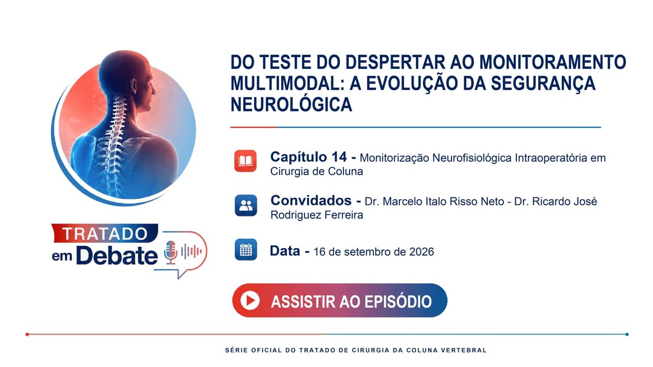
Episode 03 – Intraoperative Neurophysiological Monitoring in Spine Surgery
Videocast SBC
Watch Videocast
Treatise in Debate

Official videocast derived from the treatise chapters.

Episode 03 – Intraoperative Neurophysiological Monitoring in Spine Surgery

Scientific debate on multimodal monitoring, transcranial MEPs, SSEP, D-wave, radicular mapping, and response protocols for intraoperative alerts